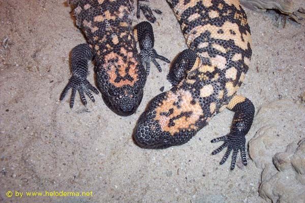

Sexes of Heloderma

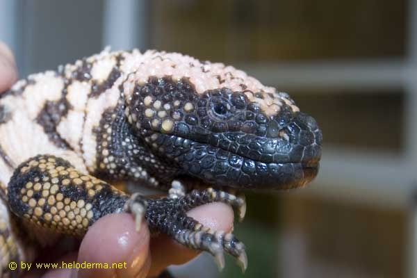
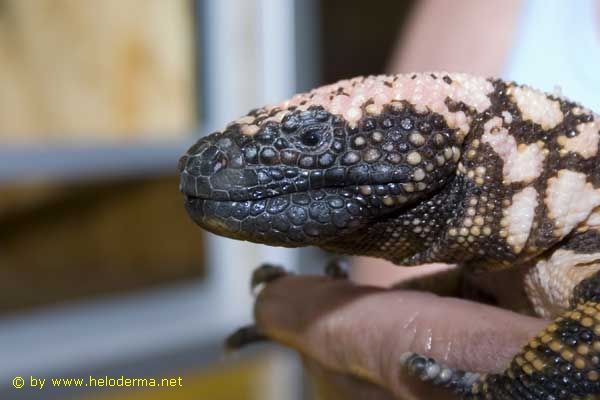
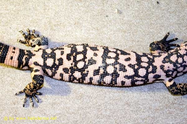
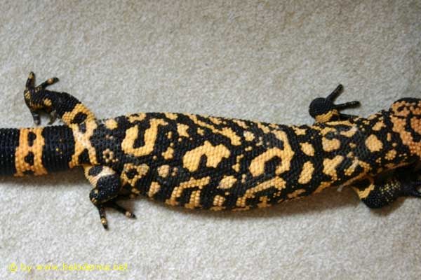
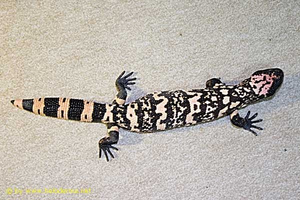
Sex determination of Heloderma suspectum proves to be difficult. Even experienced field biologists and breeders lead
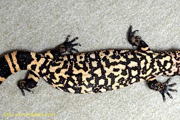
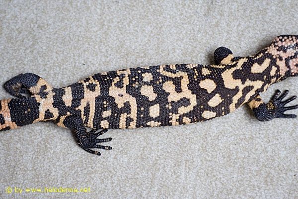
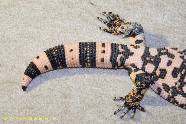
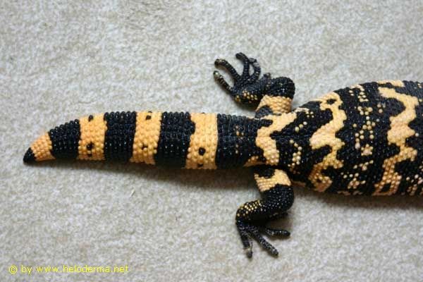
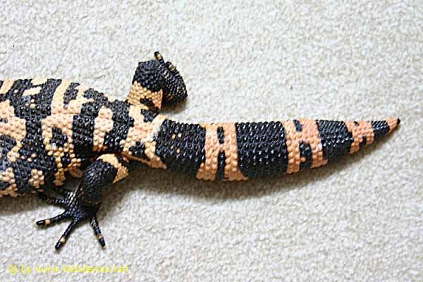
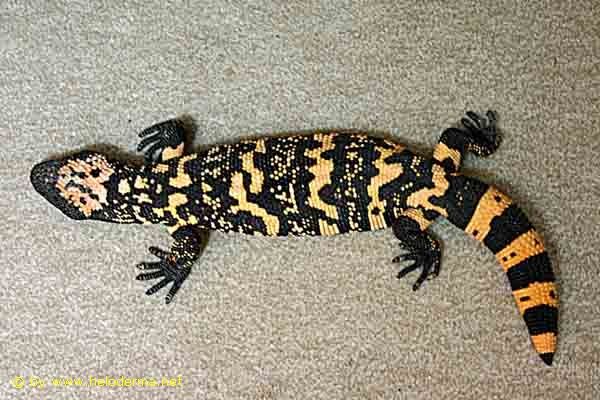
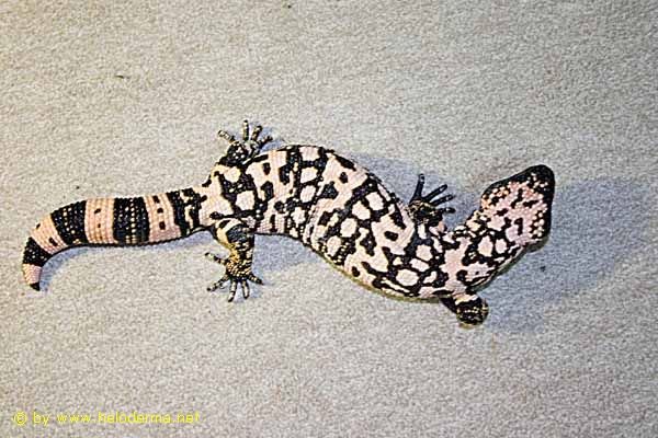
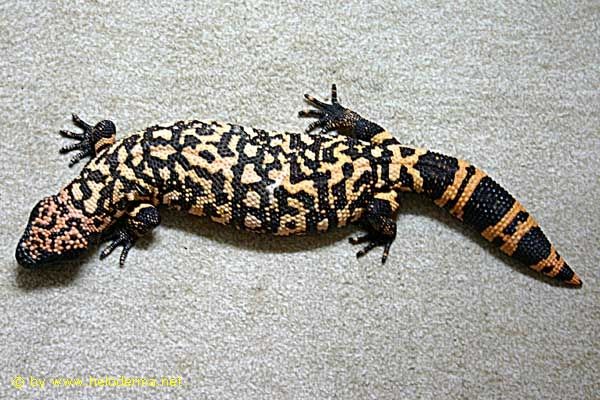
to false sex determinations (Ref. 19). Methods for sex determination in adult Heloderma:1. Body shape
|
|||||||||||||||||||||||||||||||||||||||||||||


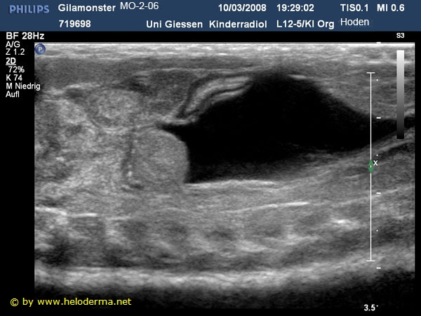
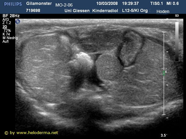
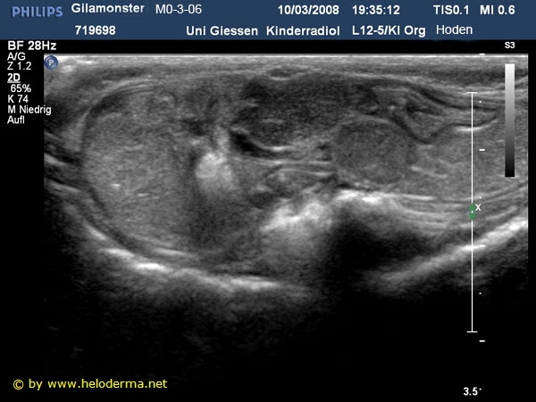
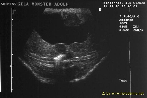
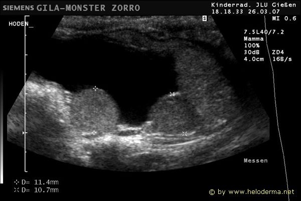
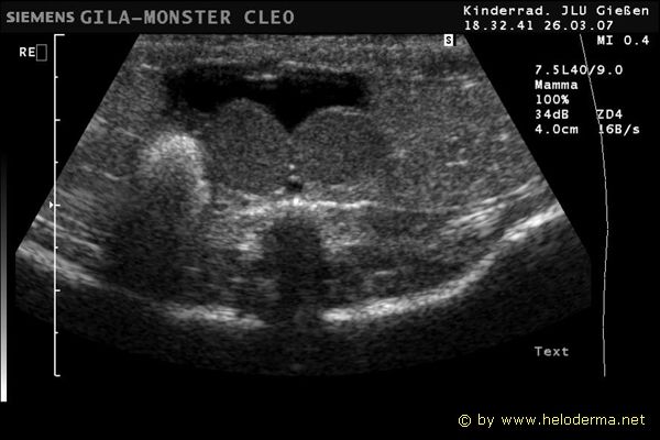
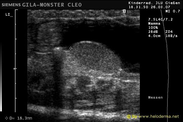
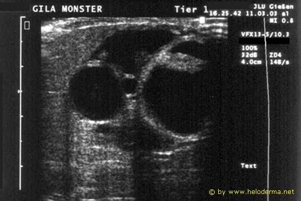
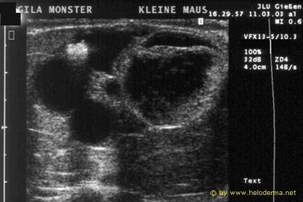
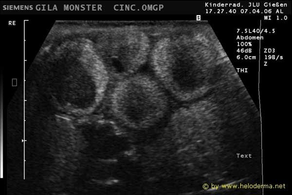
2. UltrasoundWith the help of an experienced physician, inner sexual organs such as gonads and follicles are unequivocally detectable and documented. The sexual organs are dominated by a yearly cycle. The gonads and follicles enlarge from October to March/April which is the mating season. At the end of May, these have reduced their size and functionality. Taking this into consideration, the best time to determine the sex of the specimens using the ultra-sound method would be shortly before the actual mating season (ref. 46). |
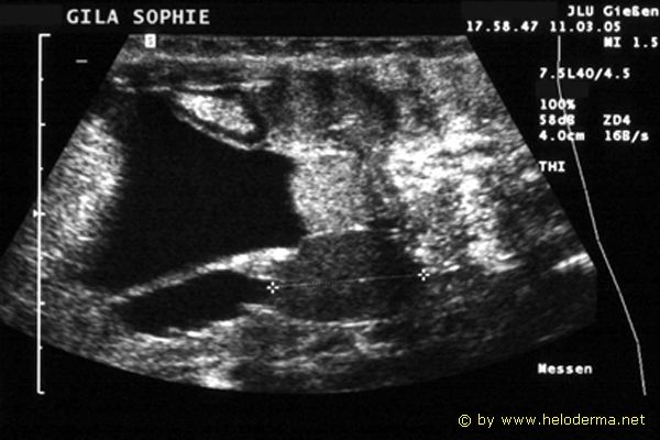
| Prints of ultrasound examinations of doubtful sexes. You can observe in text on top of the pictures that two sexes had been falsely determinated by only using body shape characteristics of the individuals: |
 |
 |
 |
 |
 |
 |
 |
 |
| >> video sequence << |
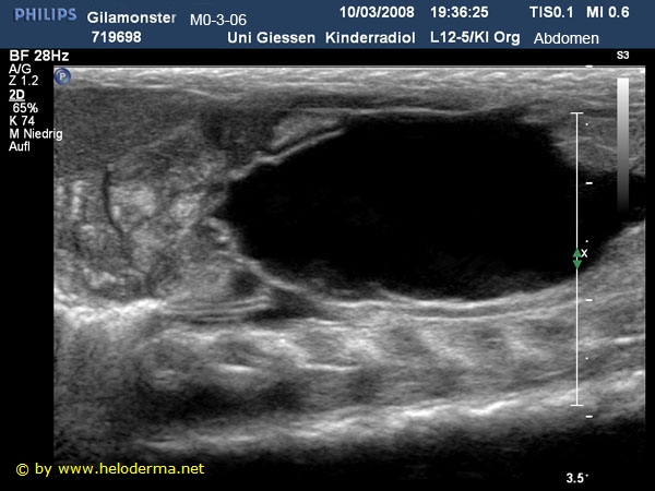
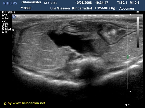
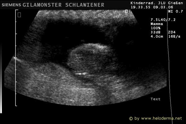
| Ultrasound examinations of sexually mature females in March, representing developing and mature follicles. |
 |
 |
 |
 |
| >> video sequence << | |||
 |
 |
 |
 |
| >> video sequence << |
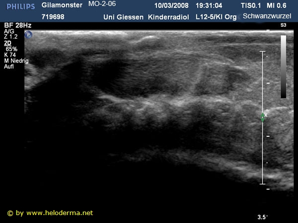
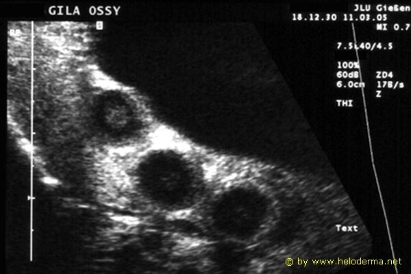
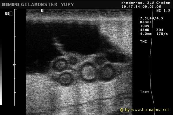
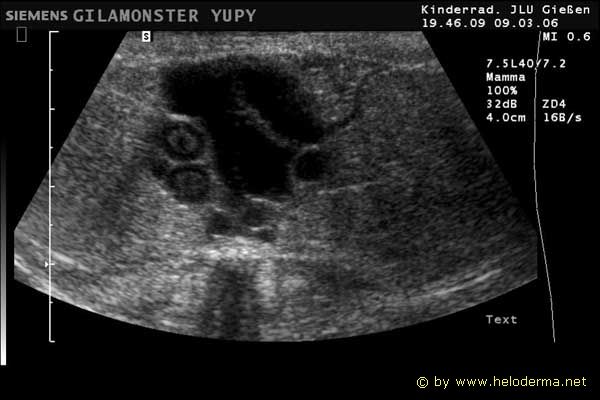
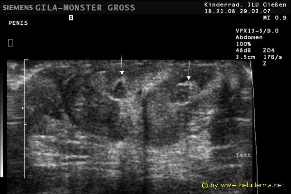
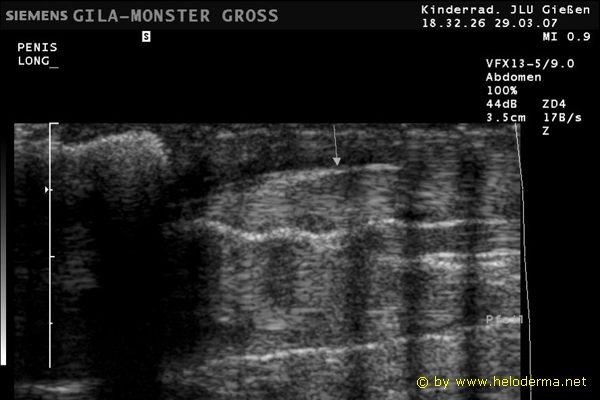
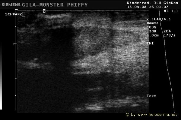
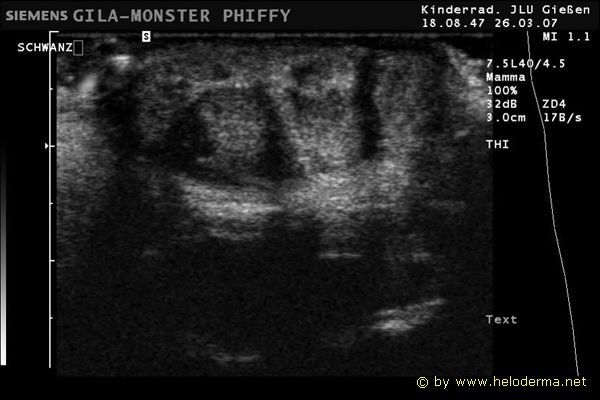
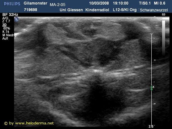
| Ultrasound examinations of adult male Heloderma suspectum in the ventral area of the tail spanning 2 cm in distance to the vent. You can observe large and variable paired soft tissue structures representing the tips of the hemipenisses with their hemipenis pockets (Ref. 35). |
 |
 |
 |
 |
|
 |
 |
 |
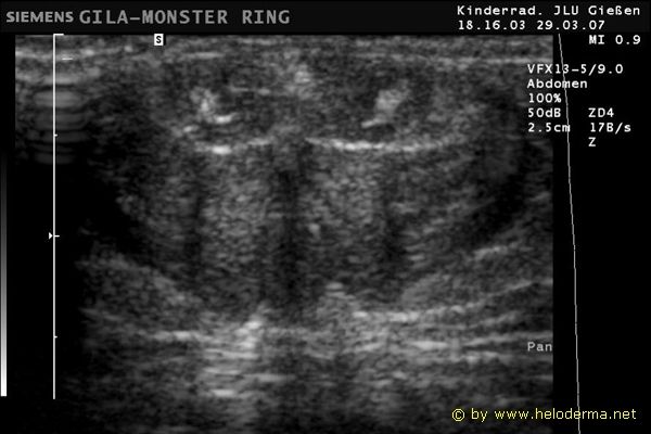 |
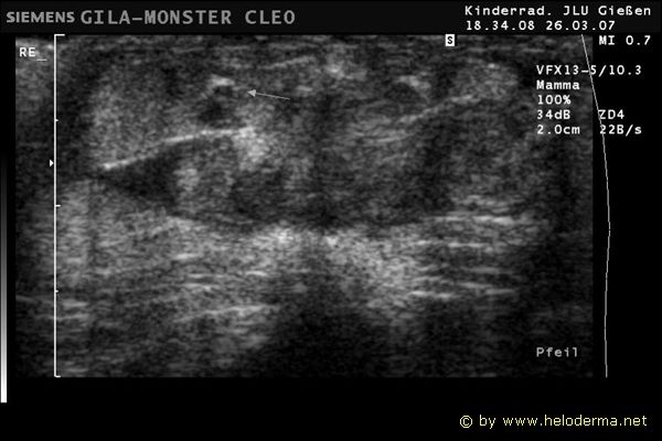
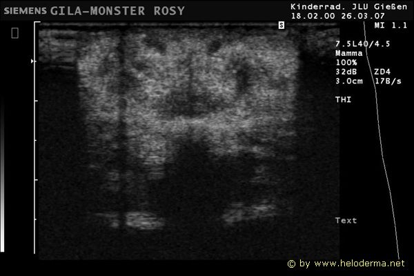
| Ultrasound examinations of adult female Heloderma suspectum in the ventral area of the tail spanning 2 cm in distance to the vent. In comparison to the male tail there is no similar structure. The tail in its full length exposes a much more consistent structure using ultrasound imaging (Ref. 35). |
 |
 |
 |
Sex determination in young Heloderma: |
|||||||||||||||||||||||||||||||||||||||||||||||
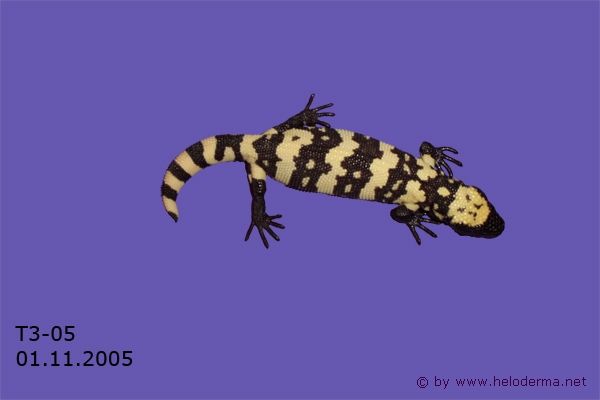
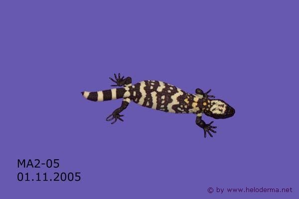
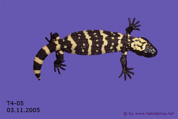
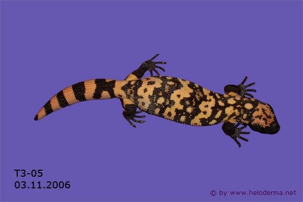
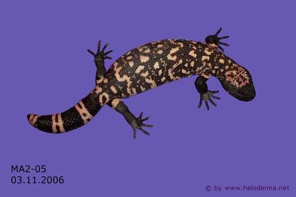
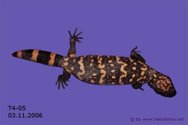
| After several consecutive breedings and observing many hatchlings, I am now of the opinion that it is possible to determine the sex in young Heloderma. Sex determination can be successful in the hatchlings when they are still filled with absorbed yolk. This makes their body shape quite distinct. After hatching, both sexes show characteristic body and head shapes. The length and shape of the tail is less useful for sex determination. | |||||||||||||||||||||||||||||||||||||||||||||||
1. Body |
|||||||||||||||||||||||||||||||||||||||||||||||
| The body of a new born male is oval shaped and appears to look bloated. The body of a female hatchling looks bloated only to her rear legs and appears more "pear-shaped". The hatchling female nevertheless has an elongated body shape. | |||||||||||||||||||||||||||||||||||||||||||||||
2. Head |
|||||||||||||||||||||||||||||||||||||||||||||||
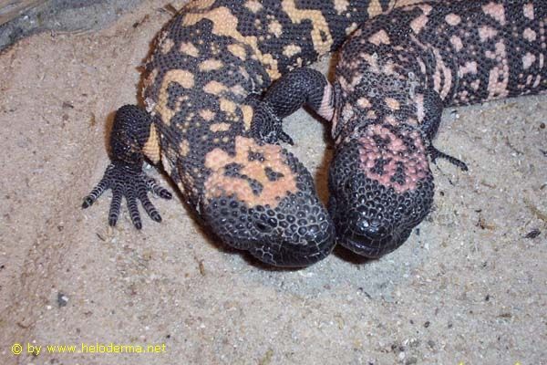
| In comparison the head of a young male Gila Monster is more triangular in shape.
This gives the impression of a larger head than that of a female. The head of a hatchling female is similar in
appearance to that of an arrowhead as it softly tapers down into the neck. Examples of male and female hatchlings from 2005:
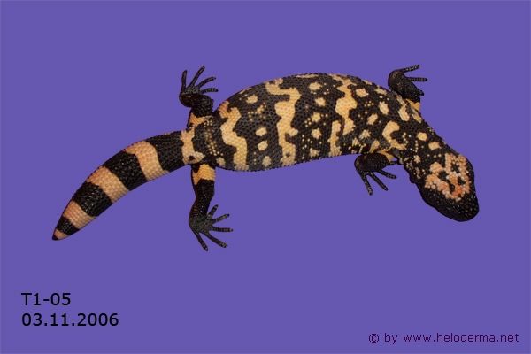
After about one month of growth, these characteristics become difficult and sex determination may not be certain. With the age of about one year young Gila Monsters show more significant body shapes for sex determination.
Comparison of head shape from the same individuals in pairs at about 12 months after hatching.
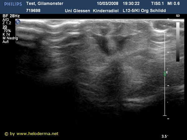
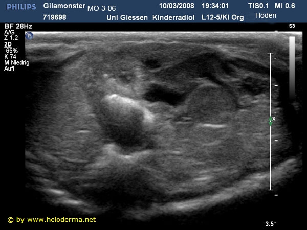
2. UltrasoundWe tried to determin the sexes of heloderma susp. from 16 months of age by ultrasound examination. The individuals came out of their first hibernation after three months and then tested. Ultrasound examination of the sexual organs and of the ventral areas of the tail of two 16 month old siblings of Heloderma suspectum.
Both specimens have a snout-vent length (SVL) of more than 230 mm and should therefore have sexual maturity (Ref. 1). In both individuals (siblings) we could detect singular roundish structures (gonade ?) , each. They are located deep in the abdominal caverty. The future will show if there will be a developement of "secondary gonades" . The two individuals came out as females. Since 2010 eggs were laid and healthy offsprings hatched.Ultrasound was performed at the Department for radiology (Prof. Dr. G. Alzen) Universitatsklinikum Giessen. Ultrasounds of the tails were performed with a 7,2 - 12,0 MHz linear ultrasound head. |
|||||||||||||||||||||||||||||||||||||||||||||||