Anatomy - Morphology

 |
| Gila - Monster |
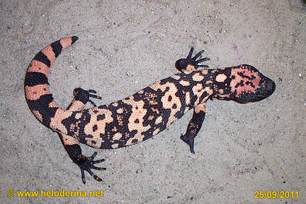
The Gila-Monster from the northern range of distribution will reach a total length of about 50 cm. Adult individuals in captivity with regular feeding can attain a weight of about 1200 g. Hatchlings weigh about 30-40 g. In the wild, individuals are significantly lower in weight, because they have less of a chance to deposit abundant fat reservoirs. The tail represents one of the fat reservoirs. After a meal, it is significantly larger in diameter for a few days. Heloderma from the southern range of distribution are naturally smaller and apparently tend to have less weight.
THE HEAD
 |
 |
 |
 |
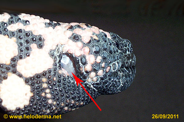
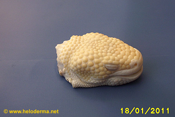
| Sense organs of the head | Pigmentation of tongue of H.susp. | External ear with tympanic membrane of H.Susp. | Organs of mouth cavity |
|
 |
 |
 |
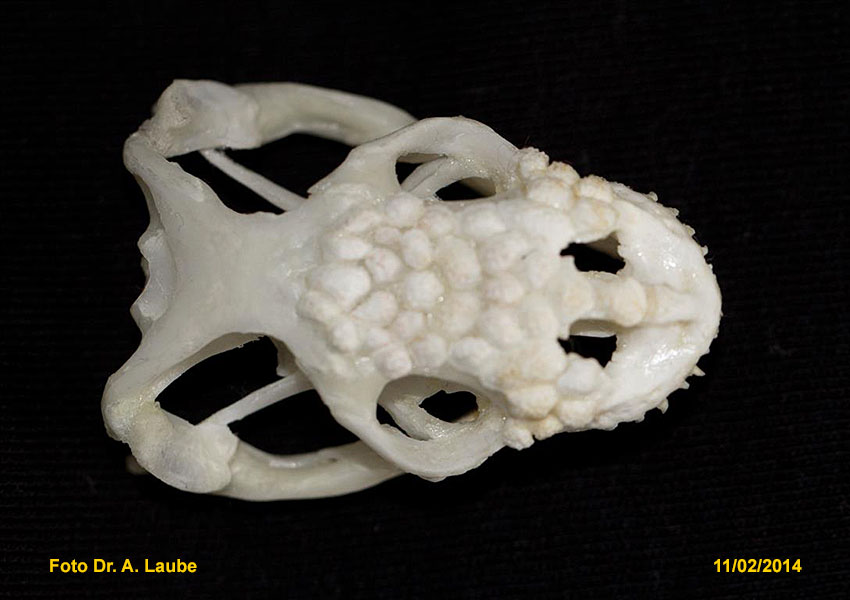
| Skull, basal view: 1) maxilla 2) palatine, 3) vomer, 4) premaxilla, 5) basioccipital bone |
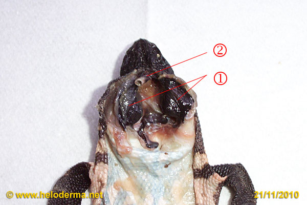
Schematics of the main muscles of the skull of H.susp.* | Exposed venom glands: 1) venom glands, 2) trachea Venom extraction (Video) |
Drawing of venom ducts (Shufeldt 1890) |
* 1) Musculus adductor (closing of jaws), 2) M. depressor mandibularis (opening of jaws), 3) M. pterygoideus (closing of jaws).
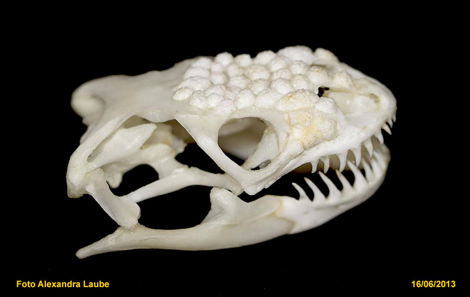
The head appears massive and stout. The osteoderms ("Skin bones") in the head region are connected to bones of the skull (Ref. 44).
The bite and holding of the victim is supported by the very strong adductor- muscle at the end of the jaws. Heloderma can easily smash the head of small rodents (pers. observ.) (Ref. 44, 46).
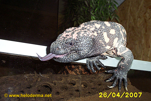
The throat of Heloderma is quite extendable. Adult lizards are capable to swallow medium sized rats, one day old chickens or juvenile rabbits.
The trachea supplies the lungs with air and oxygen. The glottis ends at about the middle of the lower part of the mouth and seals the end of the trachea. It only opens while breathing in or out (Ref. 45).
The tongue of the Heloderma is forked at its tip and is dark in color. It is fixed to the hyoid bone and is extremely flexible. The tongue serves, e.g., for food transport into the throat, drinking, swallowing, licking the content of eggs and for cleaning the outside mouth (Ref. 46).
The roof of the mouth embeds the JACOBSON-organ within small grooves.
The JACOBSON-organ analyzes smell particles which are brought via the tips of the tongue. This information is transferred to the brain to be "decoded". The JACOBSON-organ functions similar to the nose of mammals.
The ear has a strong tympanum, which is visible from the outside. Heloderma have good hearing. Soft noises at a distance of three feet are perceived and lead them to wake up. In doing so their pulse is accelerated at the side of the neck with eyes wide open.
The eye has a roundish pupil and two moveable eye lids. It has a transparent nictitating membrane (third eyelid), which can be pushed over the cornea from the nose-side angle for protection and moistening (Ref. 73).
Large, unexpected objects in motion are recognized and answered by a hissing sound.
|
 |
 |
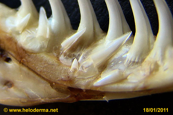 |
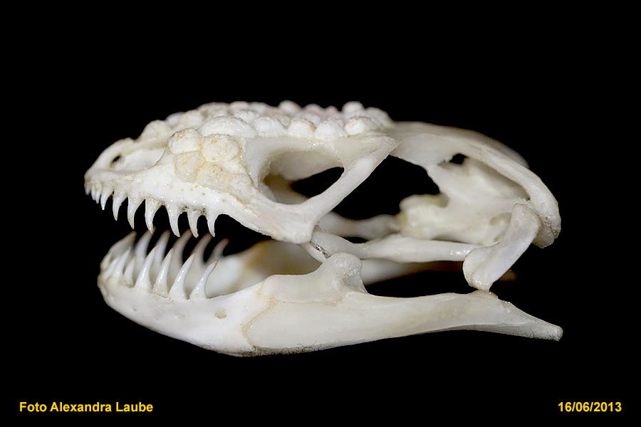
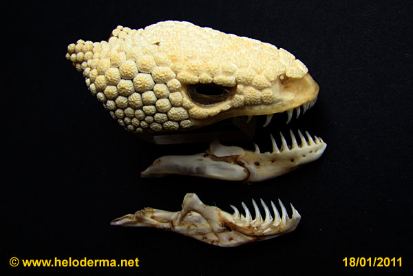
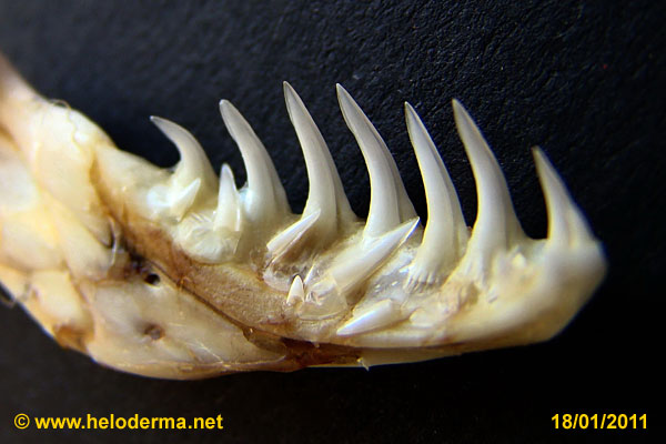
| Side view of the head of Heloderma susp. | Heloderma skull with lower jaws (mandibles) | Lower jaws with dentition | Replacement teeth at different age of development |
At both sides of the lower jaw venom glands with 3 to 4 lobes are located. These are transformed saliva glands. Via individual venom ducts of the lobes the venom is transported through the gums into the grooves of the teeth (Ref. 39). The Venom has a characteristic smell. If you smell it you can be sure the monster is irritated and ready to bite. The venom is used for protection but not to immobilize prey. (Ref. 1)
Every tooth is sharply tipped and has an apparent longitudinal groove to apply venom to a victim. The base of a tooth is flattened, irregularly grooved and tightly embedded into the beveled jaw (pleurodont).It is not possible to distinguish between incisors and molars (Ref. 39). When a tooth is replaced, its base is absorbed and the fully developed reserve tooth is brought into position from the inside of the jaw (lingual) (Ref. 39, 41, 42). The base of the new tooth is covered by new material from the jaw.
The exchange of teeth follows a wave – like pattern: at about the same time tooth No 1, 4 and 7 are replaced. The next "wave" replaces teeth No 2, 5 and 8, etc. If a tooth brakes off by accident, it has to wait until its "wave" is to start again. Heloderma change their teeth all their lives long (Ref. 46).
THE SKELETON
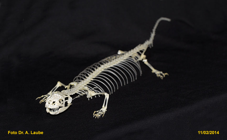 |
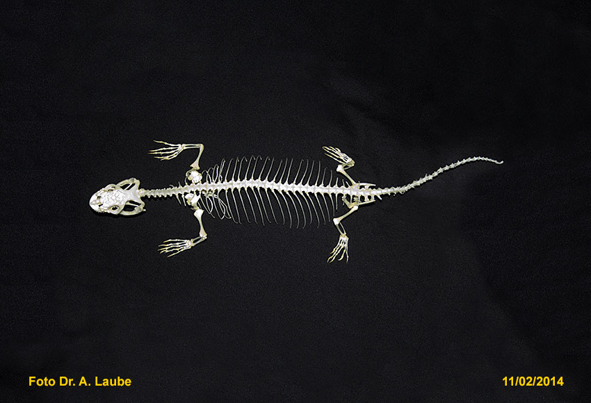
| Heloderma suspectum: complete skeleton |
 |
 |
 |
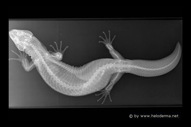 |
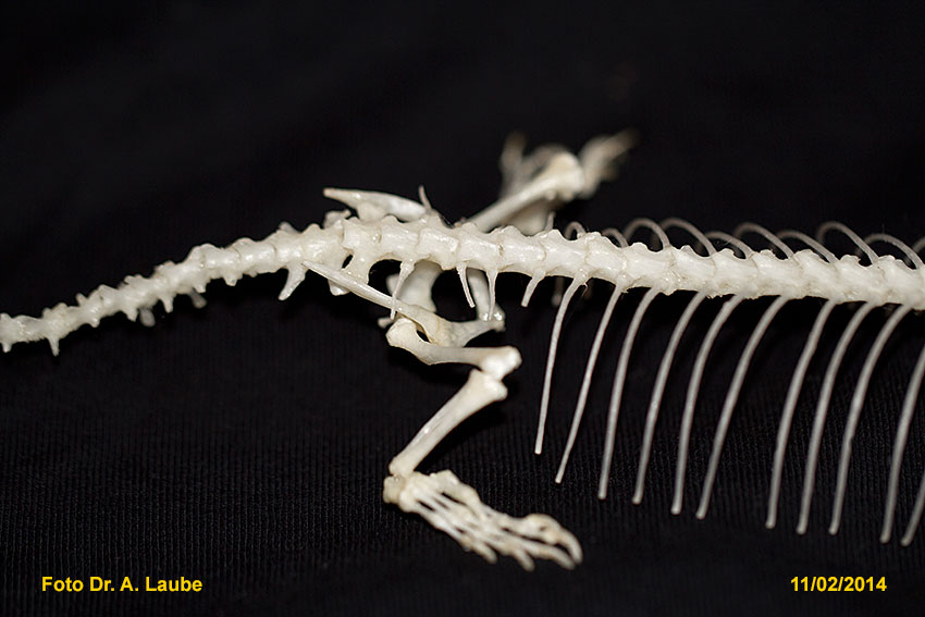
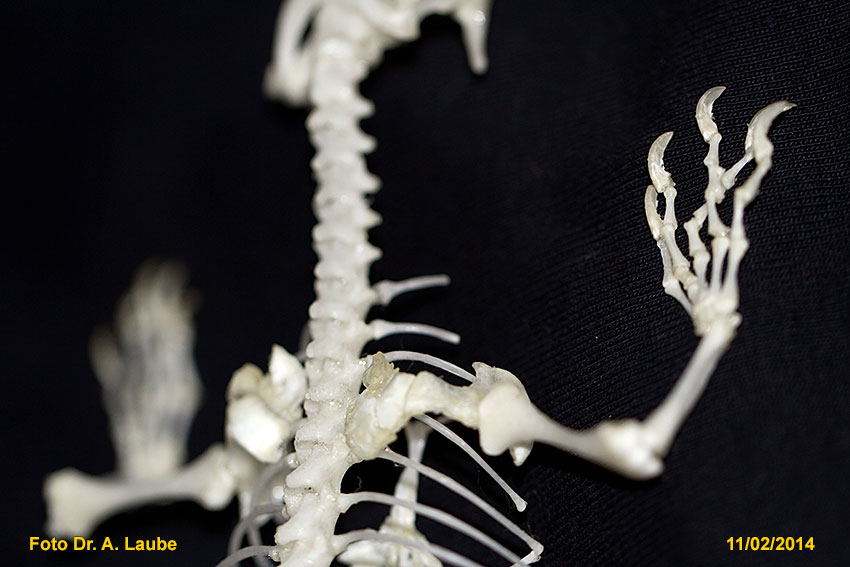
| Hello, I am a real monster - skull with front limbs | Skeleton, dorsal view | Skeleton, view from side (lateral) | X- ray of Gila monster, white dots represent osteoderms |
 |
 |
 |
 |
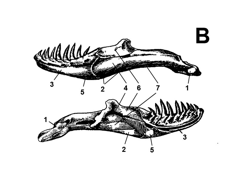
| Skull with right lower jaw (mandible), osteoderms are attached to the skull | A: Diagram of skull - view from side (lateral) | B: Diagrams of view of lower jaws (mandibles) |
Skull - top (dorsal) view |
| A: 1) Maxilla, 2) septomaxilla, 3) nasal, 4) premaxilla, 5) basioccipital, 6) supraoccipital, 7) prefrontal, 8) lacrimal, 9) jugal, 10) postfrontal, 11) frontal, 12) parietal, 13) pro- otic, 14) quadrate, 15) pterygoid, 16) ectopterygoid, 17) epipterygoid B: 1) articular, 2) angular, 3) dentary, 4) surangular, 5) splenial, 6) coronoid, 7) prearticular |
|||
 |

|
 |
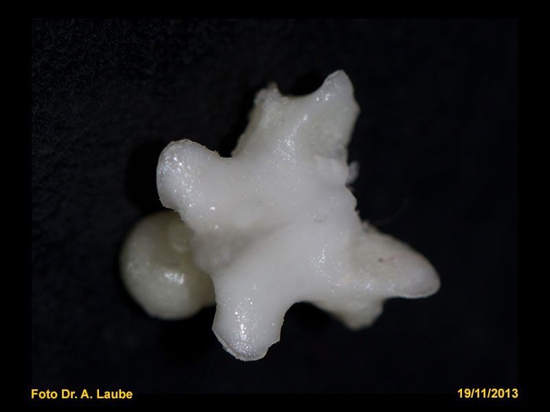 |
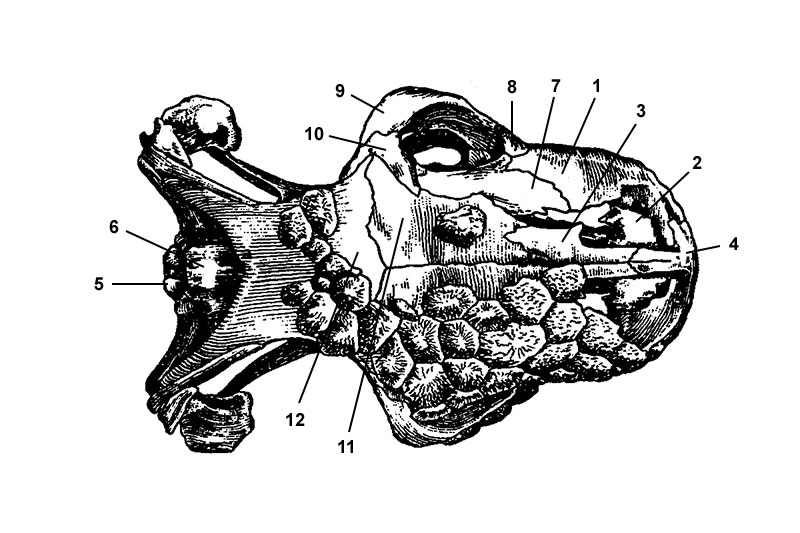
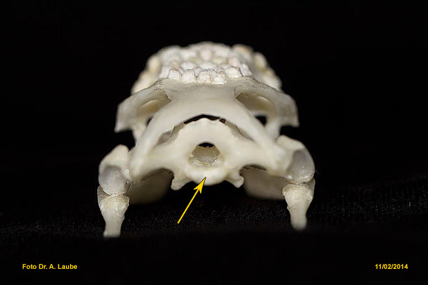
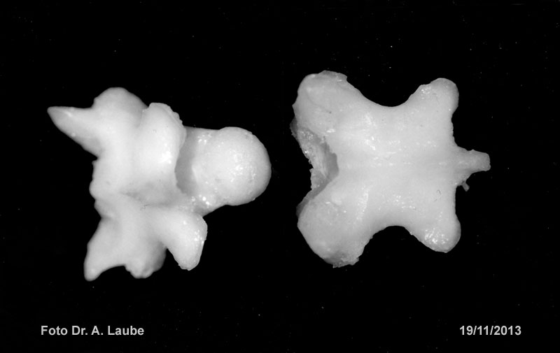
| C: Diagram of dorsal bones of the skull | D: Skull, basal view | Skull with basioccipital bone (connection to first vertebra of spinal column) | Vertebra of brest |
|
C: 1) Maxilla, 2) septomaxilla, 3) nasal, 4) premaxilla, 5) basioccipital, 6) supraoccipital, 7) prefrontal, 8) lacrimal, 9) jugal, 10) postfrontal, 11) frontal, 12) parietal D: 1) maxilla 2) palatine, 3) vomer, 4) premaxilla, 5) basioccipital bone |
|||
|
 |
 |
 |
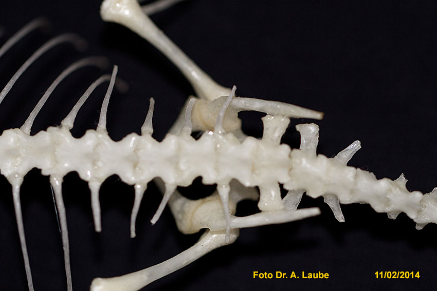
| Neighbouring vertebra | E: Diagram of top (dorsal) view of the pelvis and sacrum | Assembly of the pelvis |
Connection of thigh (femur) to pelvis |
|
E: 1) Ilium, 2) foramen for the passage of the obturator nerve, 3) pubis, 4) foramen cordiforme, 5) pectinial process, 6) ischium, 7,7a) 2 vertebrates that go to form the sacrum |
|||
 |
 |
 |
 |
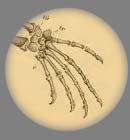
| Position of shoulder 1) scapula, 2) front arm (humerus) |
Connection of front arms to shoulder | Right front leg 1) radius, 2) upper arm (humerus), 3) ulna |
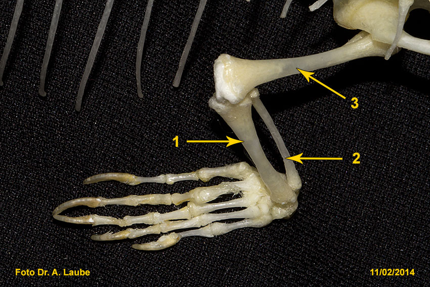
Left rear leg 1) shinbone (tibia), 2) fibula, 3) thigh (femur) |
LIMBS
The front legs of Heloderma possess strong muscles and their fingers have sharp durable claws. This enables them to dig for prey (e.g. eggs, rats, rabbits, etc.) and easily climb bushes and trees.
Their tail takes about one quarter of the total length of a specimen. Normally it tapers evenly into a sharp tip. Individuals with a roundish, stubby end of tail occur also naturally. If its tail is lost by accident it cannot be replaced (Ref. 45).
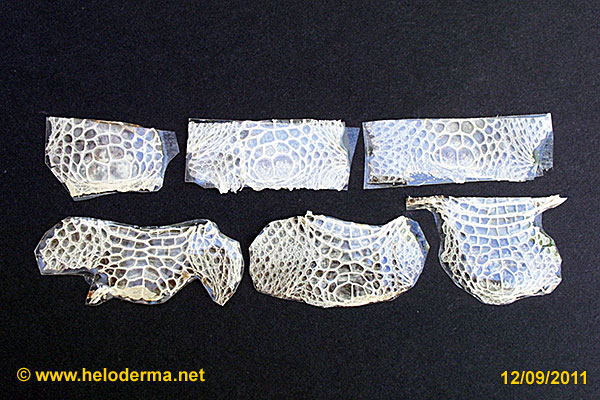
SCALES
 |
 |
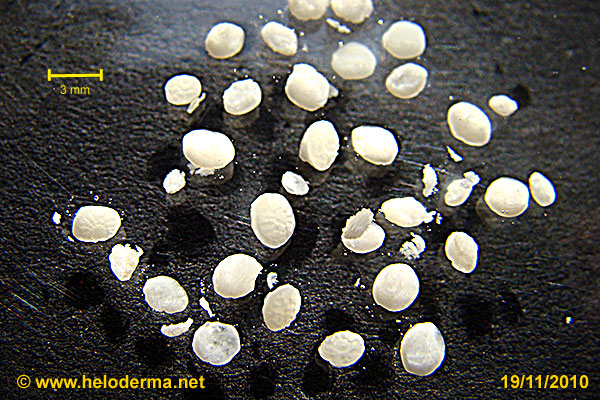 |
| Dorsal scales of H.susp. | Chains of osteoderms of dorsal scales | Different shapes of isolated osteoderms |
 |
 |
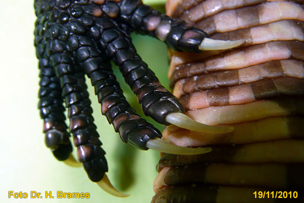 |
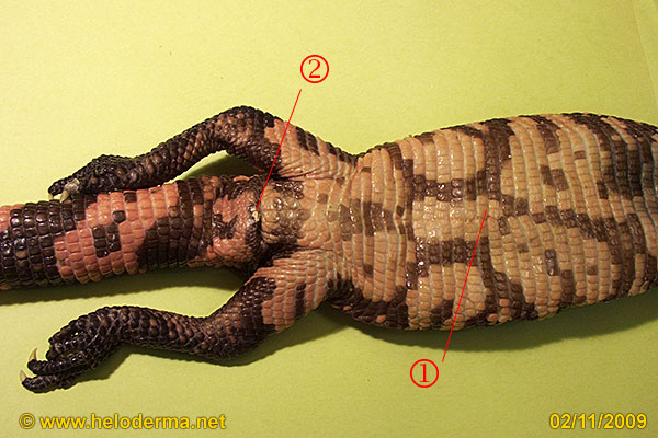
| Scales of the belly of H.susp. 1) cloaca, 2) navel | Shedded skins of cloacal region top female, bottom male | Rear right hand with claws |
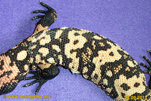
The dorsal skin of Heloderma is armored with roundish bones (osteoderms)*. When an osteoderm develops, first its tip is initiated, followed by its base and finally by the edges. In the final stage more bone material is deposited all over the structure. Osteoderms have a fibrous construction that follows the structures of the skin (cutis) (Ref. 48).
At the edges of the back towards the belly the osteoderms change their shape: while flattening they merge into a more oval shape to finally disappear within the belly scales. The ventral scales of Heloderma are flattered and show a staggered profile. The location of the entrance of the yolk sack ("naval") is to be identified all its life. Dorsal and ventral areas of different sizes and shapes of brown-black and pink to yellow are developed to form the individual patterns. The tail exposes the same colors in alternating rings (see patterns). The patterns and coloration don't give a hint to the sex of a specimen. This also holds true for the scaling of the cloacal region.
Female Heloderma start their pre-egg shed before egg disposition normally at the cloacal region. Belly and the bottom part of the tail will follow. The skin of the cloacal scales often shed in one piece.
*) Some skinks and crocodiles also have osteoderms.
INNER ORGANS
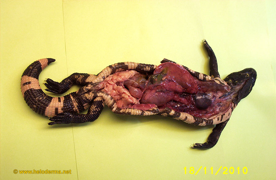 |
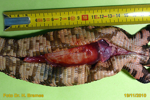
| View into a ventral opened Heloderma |
 |
 |
 |
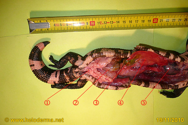
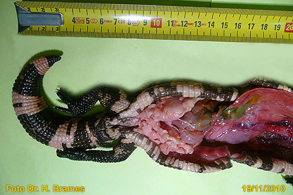
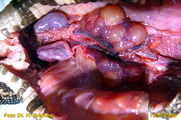
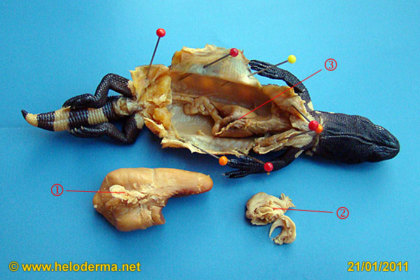
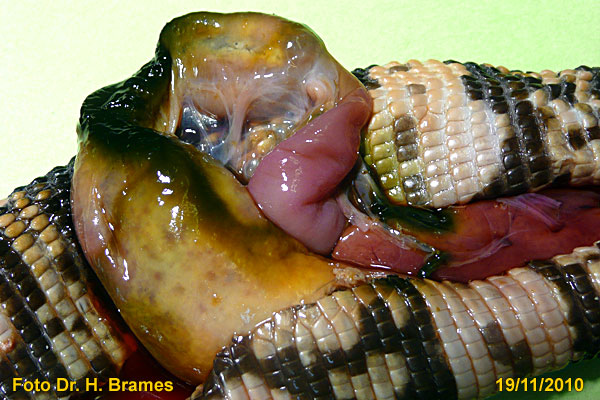
| Open ventral body: 1) fat storage, 2) small intestine, 3) gall bladder, 4) liver, 5) heart | Open ventral body | Open breast: lung, heart and liver |
 |
 |
 |
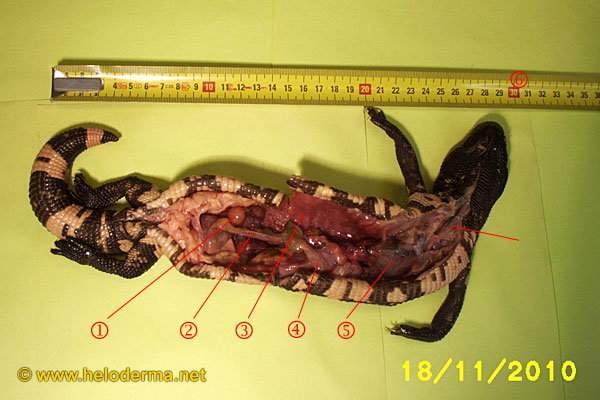
| Open Gila-Monster: 1) follicles, 2) small intestine, 3) lung, 4) stomach, 5) heart, 6) trachea |
Open Gila-Monster: 1) kidneys, 2) ovaries ; |
Open Gila-Monster: 1) kidneys, 2) follicles |
 |
 |
 |
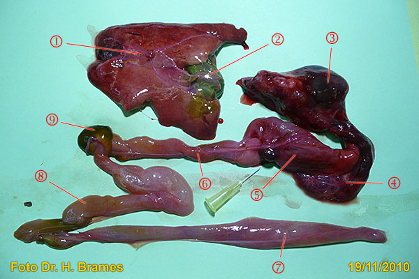
| Isolated inner organs: 1) liver, 2) gall bladder, 3) heart, 4) part of lung, 5) stomach, 6) small intestine, 7) colon, 8) intestine, 9) part of gall bladder |
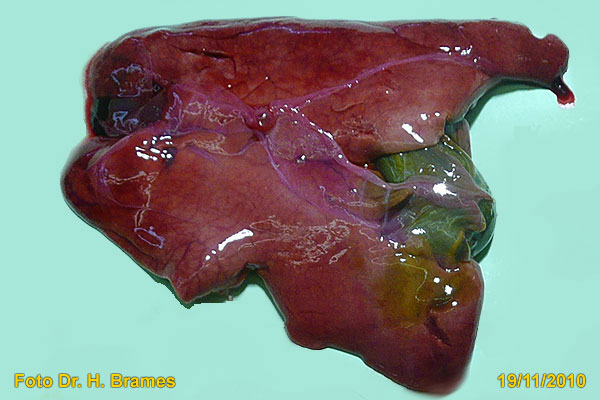
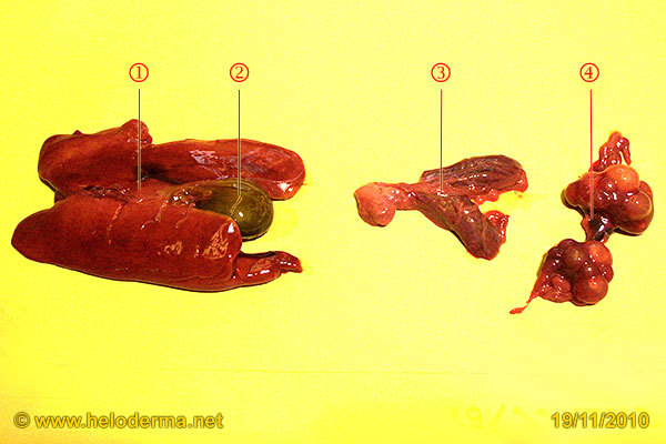
Isolated liver with gall bladder | Isolated inner organs: 1) liver, 2) gall bladder, 3) kidneys, 4) ovaries |
The heart of many lizards is equipped with two atriums, two arteries and a single ventricle (univentricular). A left and right aorta combine themselves to form the dorsal aorta. The abdominal vein runs close to the outside of the abdominal cavity (Ref. 45).
The lung of reptiles of higher biogenesis are separated from the abdominal cavity by the past pulmonary septum (no diaphragm!). They are intensively chambered and strongly expandable. This enhances their air input and reduces their breathing frequency (Ref. 46). The ability to fill the lungs with large volumes of air is used to enlarge body sizes to impress potential enemies or rivals. When a Gila-Monster feels threatened, e.g. by jealousy about prey, startled by rivals (combat!) or a female rejecting a molesting male, it will make an impressive noise. At first, it is strong hissing, that might be followed by another hissing-like noise in different rhythmic intervals. This commonly happens between individuals (pers. observ.).
To survive times of scarce food supply and for deposit of energy reserves, e.g. hibernation, the Heloderma will store fat in the kidney region and in its tail (Ref. 46).
The stomach-intestine-tract is of simple structure. A short oesophagus leads into the stomach. It is shaped like a tube and exits into the small intestine and colon for a total length together of about 20 cm (Ref. 46).
The kidneys are close to the cloaca. They don't have pyramids, no pelvises of the kidneys and no HENLE-loop, which would enable the system to reabsorb water. Nitrogen is not eliminated as urea but as poorly soluble uric acid, which is osmotically nearly neutral. Minor quantities of water can be reabsorbed from the bladder.
The urine leaves together with digested food and solid large uric acid crystals via the cloaca. Urine is not excreted via the hemipenisses – they serve "only" to transfer sperm into the female cloaca.
The reptile's liver performs similar functions to a mammalian liver. It produces proteins for the immune system and also endogenous proteins. Glucose metabolism also occurs here. It is the organ, where the provitamin D, which is formed in the skin by UV-B light (280-315 nm) is metabolized (Ref. 81) Calcidiferol, known as calcidiol (25- Hydroxycholecalciferol) or 25- Hydroxyvitamin D is a prehormon, which is formed in the liver by hydroxylation of vitamin D3 (Cholicalciferol) by the enyzyme Calciferol 25- Hydrolase. Calcifediol is then converted in the kidneys to calcetriol, a secosteroid hormone as the active form of vitamin D. Calcetriol, is then responsible for the calcium availability from the blood (e.g. for bone formation).
The gall bladder stores the gall fluid which enables the digestion of fat within the intestine. Substances of minor water solubility that cannot be excreated via the kidneys make their way by excreation through the gall bladder.
For the function and location of the sexual organs see chapter reproduction.
HATCHING PROBLEMS – DEFORMATIONS
 |
 |
 |
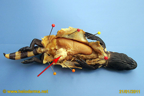
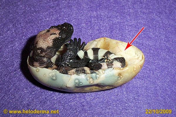
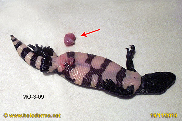
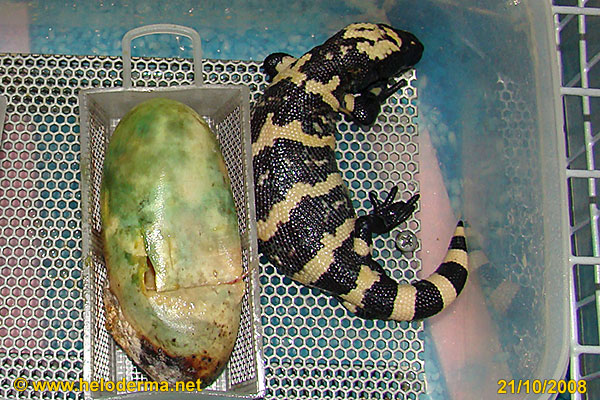
| Partially non retracted yolk sack | Anomalies: 1) deformed tail, 2) deformed spine, 3) egg shell, 4) yolk sack, 5) point of connection with shell |
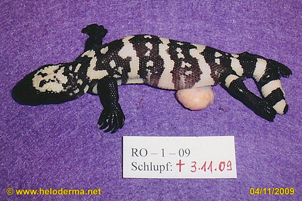
Size of yolk sack: coagulated yolk inside abdomen |
 |
 |
 |
| Sclerotic non-retractable yolk sack membrane | 1) isolated yolk sack 2) sclerotic tissue, 3) lung |
Gila-Monster with coagulated yolk within egg |
The photos show the impressive sizes of yolk sacks to be retracted into the abdomen.The depicted examples show the combination of sclerotic yolk sack membranes with coagulated, non retractable yolk contents. The ready to hatch individual is restricted and has no possibility to change its position within the egg. The egg tooth can not perform its task. The lungs cannot inflate, because the yolk sack is no longer flexible.
 |
 |
 |
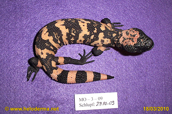
| Shortly after hatching: amputated yolk sack | Same animal with normal growth after 5 month | Heloderma: hatchling out of a badly colored egg |
Heloderma that need more than 48 hours to hatch should be carefully removed from the egg. Very often we encounter a problem with the retraction of the yolk sack. This action can be life saving for the newborn lizard. A not totally retracted yolk sack should be legated off with e.g. dental floss or similar materials. The outer part should be cut off with a pair of scissors. After 3 to 4 weeks the rest will become necrotic and fall off along with the sewing material (pers.observ.).
WARNING
By no means try to reposition any yolk sack material into the abdomen mechanically – there is a high potential of deadly infectious complications!
Operating on a female with egg retention
 |
 |
 |
 |
 |
 |
 |
 |
EXAMPLES OF DESEASES
 |
 |
 |
| Egg retention: single oversized egg | Follicles with infection | Stomach and small intestine with fungus infection |
Thanks to:
Mr. Henry Brames, vet. M.D. (Dachau) for the carry out the postmortem on several specimens of Heloderma susp. and providing some of his photographs.
Mrs. Alexandra Laube vet. M.D. (Seligenstadt) for preparing the skeleton and providing photos in total and in detail.
Mr. Jens Sievert (Berlin) for the assistance in acquiring the literature for "Anatomy".
Mr. Bernd Eidenmüller (Frankfurt) for supplying of the dead adult H. suspectum.